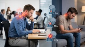
The human spine has evolved from that of 4 legged animals where the stability was very well distributed between four legs. When humans adopted the erect posture we gained the benefit of height with a better vantage to survey the surroundings,]the hands were freed for skill development. However, the weight of the whole body came to rest on the single axis of 2 legs and the centre-of-gravity shifted upwards to maintain the erect posture and the vertebrae are subjected to significant pressures.
High centre-of-gravity requires stabilization by the adductor and hamstring muscles of thigh and calf muscles to support the torso on the narrow base of the legs for even standing still. Every movement requires to be balanced putting considerable load on the lower back while walking, running and hopping. But for all that, the human back is unique in its functionality and nature ensured that this can be handled efficiently without strain on the back by readjusting the intricate muscle cover for spine and by developing lumbar thoracic and cervical spine curves, (forward convexity in the lumbar and neck regions and concavity at the thorax).
Furthermore, the paraspinal muscles are well supported by functional inter-relationships with the abdominal and lower limb muscles. A firm set of abdominal muscles share and support the load on back muscles by uniform load distribution across the oval formed by the abdominal and back muscles. The psoas muscle supporting and balancing the spine from front and sides is inserted deep into upper thigh bone.
The psoas, inner thigh muscles, hamstring and calf muscles have to be supple to maintain the correct posture and the spinal curves. Thus, to maintain a healthy spine, the muscles of not just the back but also those of abdomen and lower limbs have to be kept normal and supple and strong with regular exercise.
In the past, the life activities like physical work, sitting cross legged on the ground, squatting and walking long distances ensured that both the back and the lower limb muscles remained supple. But modern mechanized lifestyle, sitting in chairs for long hours of work, in front of TV, for dining, and use of western commode have reduced the use of these muscles except when one takes time out for exercises as an intentional part of daily schedule in which becomes more and more difficult with the demands of busy modern life.
Sedentary life style without exercises causes degenerative changes in both discs and the muscles of spine and limbs, mental stress and vitamin deficiencies play a very major role in causing back pain and development of disc issues. This is the reason for the urban epidemic of weakened vulnerable backs and painful back problems.
The spinal cord is protected by a vertebral bony canal which is almost continuous in the front with the individual vertebrae separated by intervertebral discs while at the back it forms a bony arch with a strong set of ligaments between the bones. This arrangement provides enough access for the nerves to leave and enter the spinal cord at various levels and allows a tremendous but variable amount of mobility to different parts of the spine. The arch also has paired joints at each vertebral level called facet joints.
These joints have an exquisitely sensitive inner lining called synovial membrane which has a very rich plexus of nerves to let the postural centres in the brain and spinal cord to know the exact position of the back at any given time and position. While the interverbral discs provide stability as well as mobility, the facets provide postural sensitivity (proprioceptive kinesthetic information) to accurately control back movements.
Proprioception is awareness of posture, movement and changes in equilibrium, automatic control of posture and movements, knowledge of position, weight and resistance of objects in relation to the body carried out by our nervous system at unconscious/subconcious level without us being consciously controlling these activities. Kinesthesia is the intuitive ability to perceive the extent, direction or weight of movements such as the feeling of limbs and movements so that we can walk without even looking at where we place our feet or how our body is being balanced. We need to only look at the struggles of a vertigo patient or of a patient struggling to walk in the aftermath of a any paralysis or stroke recovery to understand the vital importance of these functions
The intervertebral disc is fluid in the centre (nucleus pulposus), which is enclosed by a tough Kevlar like covering of annulus fibrosus rather like a jelly filled oval ball to buffer all the spine movements admirably. The disc has no blood supply but gets its nutrition from the surrounding tissue fluid during repetitive bending forward, backward and sideward movements.The Intervertebral disc is most relaxed in lying down position and at the end of the night it plumps up. This is the reason why back pain patients complain of worst pain in the morning which improves as they move around because the disc redistributes its fluid because of weight bearing and movements Prolonged standing, sitting in chairs and sedentary lifestyle not only weakens the disc by depriving it of its nutrition but also gradually shortens the muscles, of back inner and thigh muscles (adductors, hamstrings, quadriceps and calf muscles that control ankle position and movements).
The back muscles which have to be necessarily bulky and powerful to maintain the normal back functions, go into spasm, squeeze the vertebrae together to pressurize the discs (causing tears of annulus and prolapse) and facet joints in the arches of back causing arthralgia (painful but normal joints) in younger people, and arthritis (damaged joints) in older people with longstanding abuse of back health.
Shortened inner thigh muscles (adductors) generate stress and pain in Quadratus lumborum, erector spinae, and Psoas muscles of the opposite side (Figures ). Shortened muscles at the back thigh (hamstrings) exaggerate the normal arch of back while spasm of back muscles themselves (erector spinae and Psoas) will reduce the normal arch to straighten the lower back. All these negative influences of muscle spasms on back play a major role in causing premature aging of the lumbar spine to cause major disc problems like annular tears, prolapse etc. In certain patients who have weakened backs from birth due to minor cracks in the vertebral arches called spondylolysis, these back muscle abnormalities can result in the lumbar vertebral bodies sliding forward [anterolisthesis due to dominance of muscles in front of spine (psoas spasm)] or sliding backwards [retrolisthesis due to dominance of muscles at the back of spine (erector spinae spasm)]
Maintenance of back health: one needs to avoid sitting continuously for long hours, get up every hour and walk a little to mobilize the left and right sides of the back optimally before sitting down again. Some gentle casual stretches at this time would be ideal.
Daily yoga type flexion/extension stretches like front-to-back, (Surya Namaskar) side-to-side (Chandranamaskar), and spinal twists maintains the nutrition and health of both the discs and the back muscles toned and supple. This type of exercises prevents premature aging, spine degeneration and weakening the annulus fibrosus of the disc, which can tear and/or cause disc prolapse. The intervertebral discs, which depend on repetitive flexion/extension to draw nourishment from the surrounding fluid, are deprived of regular nutrition in people with sedentary lifestyle.
Sitting cross-legged stretches the inner thigh muscles (adductors), and Psoas muscle which runs in front of the vertebrae to the upper inner thigh. These muscles stabilize the back to optimally balance the body weight on the ischial tuberosities in the buttocks on which we sit. Sitting slouched or wedged into a fixed position in a chair for hours together without getting up plays havoc with the elasticity and suppleness of these muscles and causes tight bands to develop in these muscles to make them shorter. Shortened muscles at various levels of back and limbs pull on their tendons to cause inflammation at their ends called enthesopathy. One of the most important features of this Inflammation has 4 cardinal features of rubor (redness) dolor (pain) calor (warmth) and tumour (swelling) and in spinal problems it is this pain in the back or down the limbs that is predominant.
Low back pain (Lumbago) and radicular pain (sciatica): These are just descriptive names for the most common symptoms of back problems. These are common to those with actual disc/ facet/spondylolysis/listhesis pathology and those patients without no demonstrable pathology on X-Ray/CT scans/MRI (mechanical or idiopathic back pain) and yet other patients who have no back pathology at all but have tight muscles in the buttock called piriformis syndrome.
Mechanical or idiopathic back pain: These are patients who have back pain or radiating pain down the buttocks, back of thighs calf and outer ankle but have no vertebral, disc or facet abnormalities on MRI/CT/X-ray. There appears to be no need for back pathology to be present to produce these pains since there is no correlation between MRI findings and pain. MRI, X-rays, discography demonstrate disc anatomy but not helped predict which patients with low back pain will benefit from surgery and other interventions (Weinstein Spine 2008). Mechanical back pains exactly mimic the pains caused by disc prolapse and can be as intense in their severity and disruption of daily activities. This is because, discs facets and muscles all have specific functions to carry out in nature. The discs balance & buffer the load of body weight, Facets with their highly sensitive synovium provide the proprioceptive and kinesthetic information for streamlined perfect movements. Muscle health controls both and is in turn influenced by both. Back muscle bulk is easily 4-5 times that of bone. Spasm of these muscles can, compress the vertebrae together,
- Pressurize the discs to cause prolapse, annular tear etc.
- They also cause arthralgia of facets by compressing the surfaces of facets together to cause friction and inflammation.
- Muscles of the neck back and limbs have their own proprioceptive and kinesthetic apparatus to control posture and co-ordinate movements to the most minute detail. The intricacy of this control carried out by our muscle for normal locomotions is mind boggling in its complexity
- But most importantly, muscles have abundant pain sensors and are the main source of pain sensations at rest and movements while the sensory nervous system only serves the function of conveying these pain sensations from muscle to our brain and consciousness. Thus muscles serve the triple functions of
- Maintaining posture, involuntary proprioception, kinesthesia, and movement co-ordination.
- Carrying out conscious movements and activities AND,
- Harbour and generate pain sensations.
Most mechanical back pains are due to muscle knots called myofascial trigger points (MTrPs) and taut bands in back muscles along with muscles of thigh and calf. Similarly mechanical neck pains are due to MTrPs and taut bands in neck shoulder arm and forearm muscles. Specific treatments that address MTrPs and taut bands, the two causative problems of ALL back and neck pains can conclusively reverse these problems.
Low back and radicular pains due to actual disc/ facet/spondylolysis/listhesis pathology: These pains are labelled as discogenic (due to disc pathology) Facetogenic (due to facet pathology or combined pathologies of both discs and facets seen in spondylolysis, spondylolisthesis, lumbar canal stenosis and other pathologies like rheumatic conditions (seropositive and seronegative spondyloarthropathies like ankylosisng spondylitis), infective pathology, (tuberculosis, discitis) cancer pathology.
- direct cancer of spine or spinal cord
- metastasis to the spine from other cancers elsewhere in the body.
Back and radicular pains from piriformis syndrome: In this condition the symptoms mimic discogenic radicular pain but the discs on MRI will be normal. The reason for this is a spasm or tightening of certain muscles in the buttock in direct contact with the sciatic nerve which is sandwiched between the piriformis muscle and a trio of muscles called the superior and inferior gemelli with the obturator internus muscle in between them. When these muscles become tightened after microtears due to disuse, overuse from excessive or traumatic exercises, fall or direct trauma they cause an entrapment effect by exerting a scissor like effect on the sciatic nerve or a knife like effect when only piriformis is involved. The entrapped sciatic nerve keeps firing at the hamstring muscles in the back of thigh, the calf muscles and the shin muscles depending on which fibers of the sciatic nerve get compressed by the piriformis and/or the gemelli trio. The muscle knots formed as a result of this firing are responsible for mimicking the radicular pattern of pain in these people.
So to summarize, back and lower limb pain occur in various conditions and disc problems form only one part of that spectrum. Actually only about ….% patients with back and radicular pains have demonstrable disc pathology on MRI. In all these pains muscle is the final common expressor of pains and as such it is present in every patient with back and radicular pain. A rough representation of the causes is shown below.
- In patients of mechanical/idiopathic back pain, muscle problems are the primary and only cause of back and radicular pains.
- In a small % it is because of disc protrusions and tears A 2021 Lancet article by Knezevic etal reports that only 2-4% of back pains are due to disc pathology.
- In many, it is a mixture of disc and facet pathology, leading to formation of muscle knots which then expresses the pain. Here, muscle abnormalities are secondary to some other pathology but unless both pathologies (primary+ muscle) are addressed, the pains will not resolve.
- In rheumatic, infective and cancer pathology causing back pain, the primary pathology has to be addressed but it is only muscle treatments that will decisively relieve the pain.
The lacunae in current treatments for low back pain: The current practice in all the pain clinics across the world is to address the disc or the nerve root being pressed by the disc in disc prolapse cases, lumbar canal stenosis, chronic disc issues, spondylolisthesis by injecting steroid into the epidural space. (either the front of epidural space via a transforaminal epidural injection or back of epidural space via interlaminar epidural injections) This is followed by a prescription of pain killer and neuromodulatory medications and a referral to physical therapy. The epidural steroid injection does produce variable amount of pain relief for a variable period but eventually the pain returns when the patient attempts to become active. We believe that this is because the steroid injections treat only the neural component of pain but have no effect on the muscle knots causing the pain and various mechanical compromises in the muscle which resurface to cause pain and disability. When we do nerve blocks, we treat only half the pain but not the other more recalcitrant and persistent part of pain. This is one of the fundamental differences between the Ashirvad approach to pain and the treatments for low back pain of other pain clinics.
Ashirvad treatments for low back pain: The basic approach is that no chronic pain is limited to a nerve or a muscle or a joint, but is an amalgamation of malfunction of all the three with the brain, mind, emotion and intelligence playing a role. As such, one has to look as closely at the person with the pain as the pain itself and the possible pain generator. It has been our finding that muscle is the final common expressor of all pains whatever the initiating factor may be and in many patients it may the main pain generator as well.
So, every patient will receive some muscle treatment, be it Botox, intramuscular ozone, trigger injections with steroid or dry needling. All these treatments are guided by ultrasound visualization for specificity and accuracy and are later complemented by physiotherapy stretches. As a rule we do not start muscle strengthening exercises till the pains have come down significantly. This is because strengthening a muscle with painful MTrPs ( muscle knots) and taut bands that negatively impact many vitally important physiological functions(like coordination) is like flogging a dying horse which is incapable of getting up at this point of time. But later, when we have relaxed the taut bands and MTrPs with USGDN to relieve pain, the time is right to introduce strengthening exercises. So Ashirvad model of physiotherapy focusses on muscle stretching exercises initially and then strengthening later guided by our ultrasonography findings of muscle irritability during USGDN.
Our treatment protocol in brief addresses any primary pathology first and simultaneously address muscles
- If there is a disc prolapse annular tear, the treatment starts with an epidural steroid injection (ESI) performed with fluoroscopy guidance where real time X-ray shows out needle location in epidural space and the spread of the radioopaque dye around the nerve root being compressed. If there is a suspected facet pathology, we also do a facet injection at this time. Thereafter, all the patients will receive twice weekly USGDN sessions for the next 4-6 weeks. They also receive targeted physiotherapy. In patients with severe pain /spasm/ stiffness problems and in those with needle phobia or reluctance for needles, ultra-low dose botox injection into specific back muscles is carried out with ultrasonography visualization. For patients from outside Mumbai, botox becomes essential to complete the needling is completed within 10- 15 days.
- In elderly patients with facet arthritis, 2 consecutive test injections of local anaesthetic near the nerves that supply the facet are performed to confirm that their pain is indeed coming from arthritic facets. Once they show a positive response of >60-80% pain relief, after the test doses, radiofrequency ablation of the nerves that supply the facet is carried out. This procedure is followed by twice weekly USGDN sessions to address the muscle problems for the next 4-6 weeks. They also receive targeted physiotherapy.
- In patients with spondylolysis and listhesis, the steroid is injected into the crack in the vertebral arch called Pars interarticularis between the upper and lower articular processes of the facet. Once this inflammatory focus is addressed by the anti-inflammatory steroid injection, the secondary muscle spasm causing the pain is addressed with either Botox injection or twice weekly USGDN sessions and targeted physiotherapy.
- In infectious conditions like tuberculosis, epidural steroid injection is followed by USGDN sessions and targeted physiotherapy to address the extreme muscle spasm these patients have.
- In rheumatic conditions like seropositive and seronegative arthropathies, just USGDN sessions and targeted physiotherapy do wonders not only for pain relief but also in a significant reduction of C-reactive protein values to normal levels. Most patient go into complete remission for periods varying from a year to many years. In some patients with severe buttock and lower limb pain from sacroiliac joint involvement, one steroid injection into the sacroiliac joint will help in expediting the pain relief.
- Mechanical back pain / Idiopathic back pain – Ashirvad experience in the past 20 years has shown that this is one condition that has an exemplary response to USGDN and targeted physiothearapy along with vitamin deficiency corrections. Once complete pain relief has been achieved ( usually after 10-12 USGDN sessions) the patient has to maintain this relief with regular yoga type exercises