Last week, we had two patients with abdominal pain, which responded beautifully to Ashirvad model of pain understanding and treatment, that muscles form an integral part of every pain and need to be addressed along with the involved nerves. Or as it happened in these 2 patients just muscle treatments like Ultrasound guided Botulinum toxin (USGBTX) injection and Ultrasound guided dry needling (USGDN) proved to be all the treatment that was necessary even though both had very severe and complex pains.
For those who have not read the other blogs on this website, here is a brief explanation of our novel treatments we have developed based on a different understanding of pain. At Ashirvad Institute for Pain Management and Research, (AIPMR) we provide holistic treatment where we take detailed history and explain to patients in detail how we address all the 7 components that contribute to chronic pains such as,
1. Nervous system contribution to pain
- the nerves of the central nervous system (brain and spinal cord)
- The 12 cranial nerves that come directly from brain to supply, head, face, neck, and the whole of the internal organs from throat to anus.
- The nerves of the peripheral voluntary nervous system (the peripheral nerves that form nerve plexuses to mingle with fibers from contiguous nerves above and below and then reach the distal parts of body from top of scalp to the tips of fingers and toes
- The nerves of involuntary or autonomic nervous system (the ones that control automatic functions like breathing, circulation, digestion and most importantly muscle reflexes that control posture, muscle tone and coordination of the millions we perform so effortlessly on a daily basis.
2. Muscle contribution to pain –
At AIPMR we understand that muscles are the final common expressors of ALL CHRONIC PAINS, ALL NEUROPATHIC PAINS, (actually neuro-myo-pathic pains) and ALL VISCERAL PAINS, and not just the pains currently labelled as myofascial pains due to exercise strain and sport injuries. So, we have developed NEW Innovative therapies to address the as neuro-myopathy which is always present every chronic pain.
- Ultrasonography guided injection of Botulinum toxin (USGBTX) in ultra-low doses
- Ultrasonography guided Dry needling (USGDN)
- Ultrasonography guided trigger point injections (USGTrP)
- Ultrasonography guided Regenerative therapy (USG Regen) for injection of platelet rich plasma, stem cells etc
- Ultrasonography guided Ozone injections ( USG O3)
3. Physical therapy contribution to pain relief :
Since we understand muscles to be of primary importance, we consider all physical therapy modalities ( Yoga based exercises for stretching, strengthening of muscles, electrical modalities, Kinesio taping etc. to be an integral part of pain management
4. Counselling
Chronic Pain is probably the only disease that invariably involves the mind, feelings, mood and energy to disturb sleep, self-esteem, produce anxiety and depression. This is true for the most highly educated to the most illiterate patient, from CEOs to chaprasis. The mental contribution to anyone’s pain can only be assessed only after relieving the physical pain because in the majority of patients just pain relief will correct the mental component as well. But a little bit of support, reassurance that pain will be relieved and certain yogic practices of mindfulness and pranayama can help tremendously. For the few who need further support, referral to a clinical psychologist may be necessary
5. Correction of vitamin and mineral deficiencies is an integral part of AIPMR treatment as we believe that a deficient body cannot heal itself
6. Nutritional advice with attention to gut health
7. Referral to associated specialties like rheumatologist, diabetologist, physicians, General surgeons, neurologist and neurosurgeons, spine and orthopaedic surgeons as and when the need arises.
The 2 cases
One of the patients we saw was a 39-year-old policeman who was quite fit, used to gymming and was in peak general health. Yet, he suddenly developed such severe back pain and stiffness that he could not even move in bed. As a result, he had to be carried to one of the premier hospitals of south Mumbai. Everybody assumed that this problem must be an intervertebral disc issue but MRI showed normal disks, and no other problem in the spine. So in search of the problem they did a CT scan of the abdomen which showed a very swollen left kidney (called hydronephrosis) and a stone in the ureter that collects the urine formed in the kidney and takes it to the urinary bladder. Later history revealed that he had had ureteric stones, twice before in the past 6 years. So everyone was relieved that an answer had been found, and he was transferred from a spine specialist to the urologist.
The swollen kidney (hydronephrosis) was treated with what is called as a double J (DJ) stent, a thin coiled tube inserted from the upper end of ureter to the urinary bladder so that the urine can be drained out of the swollen kidney for a period of time allowing the kidney to recover. The stone is usually left alone at this time to be removed later along with the DJ stent once the kidney has recovered. However, despite kidney being decompressed successfully, this patient continued to have severe back pain and stiffness, and he was unable to get up or walk or help himself despite the strong pain killer injections that he was receiving. This was a difficult situation for his family and friends. Then one of his friends, another surgeon in the same hospital who had been treated at AIPMR, called me to ask if the patient could be helped.
I explained that this was likely to be the neuromyopathic expression of the stone and advised that he be given a neuromodulator medication pregabalin (lyrica ®) in addition to the pain killers. This neuromodulator medication which works on the nerves and indirectly relaxes muscle (AIPMR understanding not mentioned in the drug information) made a big difference allowing him to sleep comfortably but the next day when he came to the clinic, we found that he had difficulty getting up from a chair and could only walk very slowly, gingerly despite the painkillers he had received just prior to discharge and could not bend at all. (not even 50 movement)
I explained to him that this problem was because his back muscles had stiffened in response to the stretching of the capsule of the kidney by the backpressure of hydronephrosis and the irritation of ureter by the kidney stone. The patient understood this, but I could see that he thought it was long shot. Then I further explained to him that the internal organs are voiceless to express/convey their distress directly to the brain and mind but use a natural process called ‘Pain referral’ to the muscles of the back and abdomen to reflect their pain which then reaches the brain. This is the pathway nature uses to convey problems in the gut or the genitourinary system to the brain/mind so that we become aware of the pain. This process is called viscero-somatic convergence. (internal organ muscle spasm with knot formation musculosensory nerves Brain).
This happens because during development of the baby’s body inside its mother’s womb the nerves to the gut, the genito- urinary system and the muscles of body wall receive nerve supply from the same segments of the spinal cord, usually between thoracic 5 to thoracic 11 and in the case of ureter, it goes down till about Lumbar two and three levels(L1- L2). When the ureteric stone is passing down from the kidney to the bladder, the patients get severe flank pain. (flank is the side of the abdomen going down to the groin) Our Patient had been having this problem of muscle stiffening over the years with repeated stone formation, but he had not experienced severe pain because the spasm of the muscles had not reached a threshold above which the body perceives it as Pain. Nature has designed the human body in such a way that “the show must go on” so that we keep functioning normally even though the situation inside is abnormal. Muscle spasm and myofascial trigger points are like an iceberg which keeps growing below the water surface but becomes visible only once it grows big enough to be seen above the water, making pain the tip of the iceberg of abnormality.
So, though he had had stones and even taken lithotripsy treatment to shatter the stones once before, he had not experienced the typical severe pain travelling from the back to the groin area. Apparently, this time round, his muscle spasm had reached above and beyond the pain threshold because the stone was blocking the ureter causing back pressure from the urine collection on the kidney gradually inflating it and stretching the capsule. Unfortunately, this episode had become extremely painful for him.
After this long explanation. He understood the problem and then wanted the treatment for releasing the muscle spasm, but he had a doubt. Why had his other doctors not told him about this problem or the connection between the internal organs and the muscles? I then explained to him that some of these ideas have evolved at AIPMR by interaction with previous patients in whom these treatments had produced good results. His problem and its severity were uncommon considering the number of patients who get urinary stones but who recover uneventfully after urology treatment. In medicine new ideas take time for assimilation and require proof and validation via scientific publications, lectures and interaction between different specialists. Also, every specialty tends to publish in journals which are read within their own specialty. Pain is the symptom that straddles many specialties and specialists from other specialties need to accept pain management and interact with pain specialists more for a holistic management of unusual problems. It is only when the pain persists despite the stone removal that they get puzzled and then seek help from a Pain Specialist. He had reached me because of his friend who was aware of AIPMR treatments.
The patient agreed to the treatment plan of USGBTX into various muscle layers of the back and abdomen with ultrasonography guidance, followed by another treatment called ultrasonography, guided dry needling. (USGDN) which is needed to finetune the effects of USGBTX, which has to be injected in ultra-low doses to avoid weakening of back muscles. The residual spasm that remains after ultra-low dose USGBTX gets relieved by the USGDN, which is done in a very systematic way to completely relax each and every muscle involved in the pain. (see our blog on Muscle, the blind spot of chronic pain for more detailed information)
Immediately after the USGBTX (which takes about 2-3 days to act) followed by the first session of USGDN to provide immediate relief, his pain and stiffness was very much relieved. When he returned three days later for his 2nd USGDN session, he had been sleeping well, was quite comfortable with daily routine and could do quite good physical therapy and wanted to know if he could return to work. I told him that he could return to work, but not to get worried if the increased activity caused some return of pain because it could be addressed quite easily with repeat sessions of USGDN. He was quite okay with that and started going back to work, and he did come back for 2more repeat sessions of needling and is doing well. However, the kidney stone is still there inside and till it is removed his pains could act up but in a much reduced level, he knows that even after the stone removal, he has to undergo to 3 sessions of USGDN to eliminate the residual effects of the ureteric irritation by the stone on his back muscles, but once the stone is removed, and residual muscle knots resolved, his pains will not return.
In addition to USGBTX, USGDN and physical therapy, he needed correction of his vitamin deficiencies which were significant and probably had contributed to the severity of his pain. He was given injections of vitamins D and B12 and was also advised magnesium supplements. He had no significant mental contribution to the pain so there was no need to address that. After the stone removal and analysis, he will receive some dietary advice to avoid certain foods which might make him prone for repeat stone formation and of course adequate hydration. As a gym Person he was anxious to restart but has been advised to start low and go slow till the USGDN sessions after the stone removal. Thereafter he should have no need for any pain treatment.
Patient no 2
The second Patient was seen in Nair Hosp pain clinic by Drs Varsha Suryavanshi and Bhaskar Patil who have trained at AIPMR and follow the same treatment protocol.
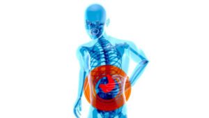
This was a little girl of 11 years who had chronic pancreatitis. Pancreas is an organ inside our abdomen behind the stomach in the left half of upper abdomen. It is the organ that produces lot of digestive enzymes and also insulin. When these digestive enzymes breach the protective mechanisms and reach the abdominal cavity instead of into the gut, it causes pancreatitis. It is called acute pancreatitis at the first instance when it presents with severe pain but later settles down. But in a variable % of patients (6.4-36%) it can become what is called as chronic pancreatitis. The reported annual incidence in US is 0.5-2 cases per 100,000 children.
This child had this ever since she was about nine years and now she is 11, which means she had two years of pain, but it had been manageable with pain killers at home but had worsened in the last six months when it stopped being responsive to pain medicines and specific treatment for chronic pancreatitis. In the last 3 weeks, she had required admission to the hospital with intravenous pain killers to control the pain but which could only provide partial relief for just 2-3 hours.
They had done what is called as endoscopic retrograde cholangiopancreatography (ERCP), which is a procedure which is done through the gastroscopy to enter the pancreatic duct and demonstrate any blockages and clear the pathway for the juices to flow out. This had shown chronic atrophic pancreatitis and had inserted a stent in the pancreatic duct. In spite of this procedure, the pain was still persisting. The pain in fact had become worse in this child, and she was unable to sit even for more than 2-3 minutes. She was unable to stand or walk, and she was obviously not able to eat with all these problems with repetitive vomiting. The whole family. I was very much distressed because of small little girl in so much pain which affects not just the sufferer but also the whole family.
They were sad and helpless because they had gone to private clinics, hospitals and then they had reached Nair Hospital where senior gastroenterologists had done a definitive treatment for the pancreatitis, that of pancreatic duct stenting but they had not been able to relieve the pain, which is why she was referred to the Pain clinic. People were trained at AIPMR are very much aware of viscero-somatic convergence, the pain referral to back and abdomen and that the pain can be treated by relaxing the muscles of the back and as I mentioned in the previous case of the policeman. Pancreas is an internal organ and during its development in the fetal period, (as the baby is being formed) it shares its nerve supply with the muscles of abdomen and back. During attacks of pancreatitis, when the pancreatic enzymes are being released into the abdomen when viscero-somatic convergence becomes operative to produce fresh waves of muscle spasm and pain till it goes well beyond the threshold of pain, and this pain persists, even though the original cause may be remedied by definitive treatment by gastroenterologists who had done stenting of pancreatic duct to ensure a free flow of enzymes safely inside the gut instead of causing back pressure in the pancreas.
The enzyme levels had already shoed a decrease showing that the problem had been remedied by the ERCP stenting procedure. Pain still continues because the initial waves of muscle spasm which are caused by the pancreatitis, generates what is called as myofascial trigger points (MTrPs) or knots in the muscles of the abdomen and back. Once formed, these muscles knots do not resolve by themselves nor by the gastroenterology treatments. They persist even if the pancreatitis is cured and need to be actively treated. This is the reason why the child continued to be in pain even though the actual problem had resolved. This is a situation which is very difficult for most doctors even to understand the mechanism of pain even though they understand the problem which gives rise to the pain and they can treat it well with stents or surgery with the simple equation that there is a disease.
The disease is gone after their treatment so the pain which is a pointer to the disease should go. But Pain behaves in a very aberrant manner. Chronic pain, especially has its own behaviour pattern which most doctors are just discovering. We have published a paper on this in pancreatic cancer and also about another urological problem called interstitial cystitis in other journals. That the connection between the organ problem and pain lies in muscle. The muscle spasm which leads to myofascial triggers or muscle knots, which proceed to cause pain and these are independent of the original problem that gave rise to them in the first place.
With each recurring episode of pancreatic problem, the muscles get tighter and tighter, and more and more painful so this child had to be brought to the Pain clinic in a wheelchair, and she had to be shifted onto the bed by her father. But once Dr Varsha did the needling of the back in a very specific manner under ultrasound guidance which hit the pain problem spot on. Ultrasonography shows very clearly which muscle is where and also shows certain events in the muscle which sigle out the muscles with MTrPs.
It helps to safeguard the underlying organs because we can see where the organs are how deep they are, and the needles are inserted above only into the muscles with perfect visualization. So she did this kind of needling into the abdominal muscles, and the three components of the back muscle called Erector spinae, another muscle called quadratus lumborum and yet another muscle, called psoas which is very deep inside, and it cannot be visualized without expert ultrasonography. But once we can look through the ultrasonography, we can very easily introduce needles accurately into these muscles.
Of course, it takes quite a lot of learning curve and skill to develop these abilities, but AIPMR specializes in this training so it can be done and is being practiced across India by the people who trained at AIPMR. Needling done in this manner relieved the child so much that she says that her pain was zero just after one USGDN session and the girl who had come on a wheelchair could walk briskly immediately after this session. Of course, the father and the child were told that they would have to come for another at least 4-5 sessions because these problems do not get settled with only one session of needling. The number of muscle knots which are formed over months, years (In this child, two years), will be significant and in considerable numbers so unless all of them are eliminated, the pains will recur.
This child has had about three sessions. She is eating well, continues to be completely pain-free and is now smiling all the time instead of crying. So, the father was very very thankful, and said “we did not know what to do because we have gone to the so many doctors, nursing homes, specialists and hospitals but if specialists can’t solve this problem, where can we go? What can we do? I am very thankful to the Nair Hospital Pain clinic for identifying this problem and treating it”.
Focusing on muscle pathology and judicious use of USGBTX and USGDN can produce very dramatic answers to many confounding and difficult googly situations in chronic pain.